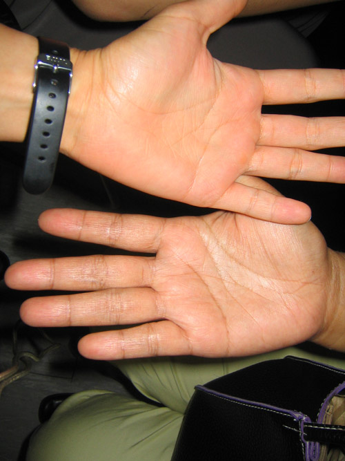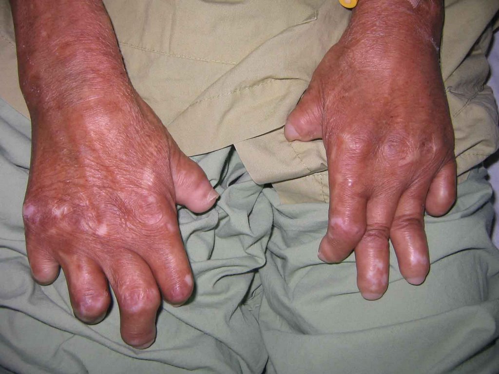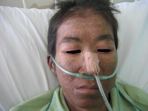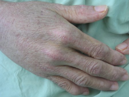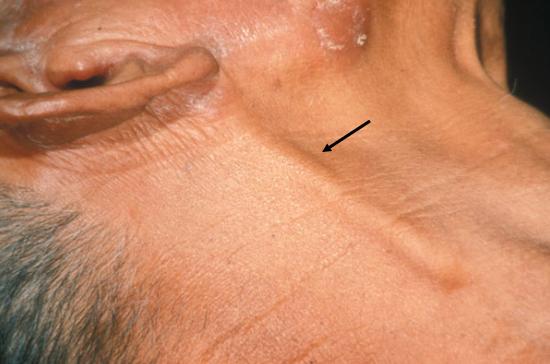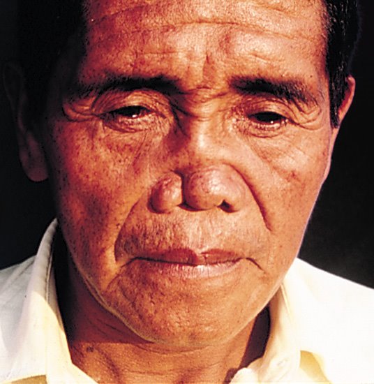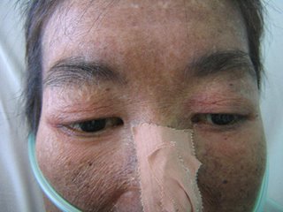
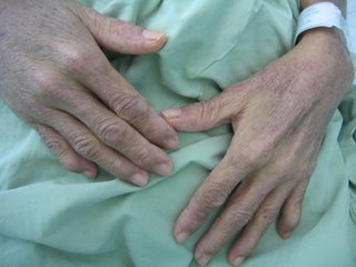
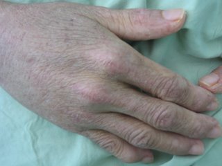
This lady has dermatomyositis. She has a heliotrope rash over the knuckles (GOTTRON"S Papules), back of the hands, eyelids and periorbital area. She also has nail fold telangiectasia.
(Look also in elbows and knees for Gottron's papules, it may be the only clue you have)
She also has Cushiongoid features, likely due to steroid treatment. (Mention other complications of steroid treatment if any-Cataract, vertebrae fracture, obesity).
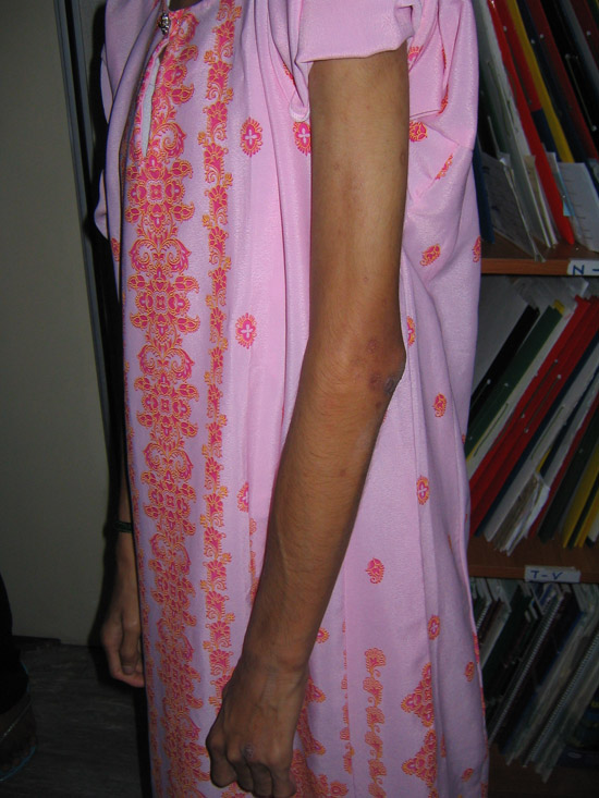
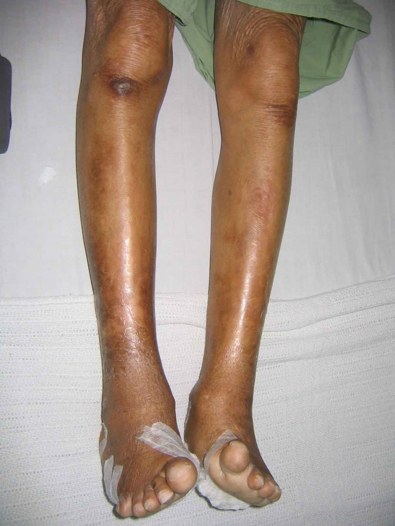
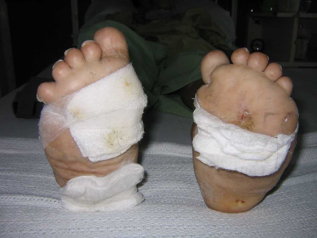
There is also proximal muscle weakness and tenderness and the patient has had a muscle biopsy done (look for scars on thigh/upper arm)
Functionally, she has impaired swallowing as she requires a Nasogastric tube
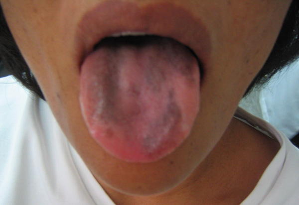
NB Candidates often mispronouce "Gottron's" as "Grotton's". Be careful! Look for other things such as Raynauds, SLE
Questions that could be asked
1. How would you manage this patient?
**I would like to confirm my diagnosis of dermatomyositis by doing the appropriate investigations (eg muscle biopsy, Electromyelogram)
**As this lady is over 40, I would also investigate for underlying malignancy (mention NPC, breast, ovary, Lung and GIT) I would also investigate for connective tissue diseases.
**I will also start patient on high dose steroids and add on steroid sparing drugs.This patient also needs nutritional support as she is cachetic and has dysphagia caused by the disease.(Hopefully by the time you reach this station, your time is up)
2. How to differentiate this rash from SLE rash?
**In SLE, the rash spares the knuckles, and involves the phalanges.
3. How would you classify polymyositis-dermatomyositi?
**I Primary idiopathic polymyositis
**II Primary idiopathic dermatomyositis
**III Dermatomyositis (Or polymyositis) with neoplasm
**IV Childhood dermatomyositis
**V polymyositis/dermatomyositis with collagen vascular disease
The above case and photos are contributed by Giant Eagle
Labels: Skin, Station 5


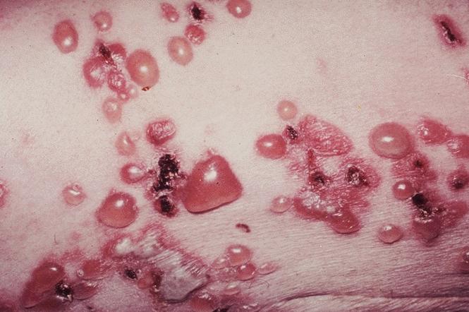
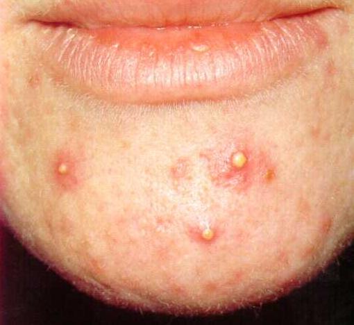
 This gentleman has multiple similar lesions distributed over face, limbs as well as trunk. Please describe the lesions.