PACES MRCP UK - Where MRCPians Meet Since 2006

MRCP is well establised as an entry exam for advanced specialist training in many countries including Malaysia. It consists of 3 paper i.e. Part1, Part2(written tests) and PACES. PACES in full means Practical Assessment of Clinical Examination Skills. It is the third part and the candidate is assessed by fellows of RCP. I passed my PACES in 2005. I am glad that many seniors had guided me throughout my preparation for PACES and I wish to share my experiences with PACES candidates via this blog.


Wednesday, June 13, 2007

Respiratory Quiz


During yesterday’s MRCP mock exam, Wuchereria had a tough time with one of the local examiner.

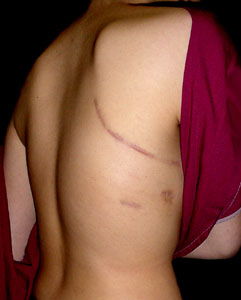
This patient has clubbing with cyanosis and bilateral fine crepitations over the bases with a scar as seen in the picture. He also has an area of consolidation over the R lower zone

Examiner : How do you correlate the scar and the findings ?

Any takers?

This case is contributed by Wuchereria.

Labels: ,

Monday, June 11, 2007

SVC Obstruction


There is prominent veins over this patient's anterior chest wall. They are tortuous and dilated. The neck veins are engorged with loss of pulsation. The face looks plethoric and suffused.

This is superior vena cava obstruction.

This patient has undergone recent radiotherapy for this medical emergency condition as evidenced by fresh radiation ink marks and skin erythema over the same area.

Any other signs to mention? How do you tie them up to form a complete diagnosis?

What are the other clinical clues to the unifying diagnosis?

Photo courtesy of Wuchereria

Labels: ,

Thursday, April 20, 2006

Fibrosing Alveolitis

The respiratory case that I had in UK was rheumatoid lung disease. I got no problem in detecting the clubbing and fine inspiratory crepitations bibasally. But, the outcome was 2/2!

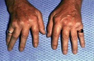
Pitfall: I failed to identify the underlying cause for fibrosing alveolitis. I missed out the obvious sign i.e. Rheumatoid hand changes!

The moral of the story to learn is ALWAYS look for the underlying cause for fibroising alveolitis as cryptogenic fibrosing alveolitis is RARE for PACES!

Face: malar rash (SLE)
telangiectasia, beak nose, microstomia (scleroderma)
facial pigmentation (amiodarone)
heliotrope rash (dermatomyositis)
Hand: RA, sclerodactyly

Never miss RA hands in respiratory station!

Labels:

Sunday, April 16, 2006

Crepitations and Clubbing


Record :
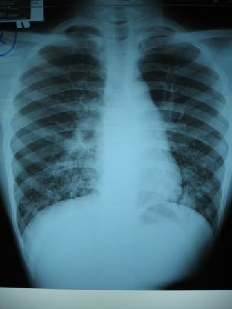
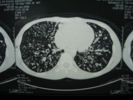
This young boy is tachypnoic at rest with a RR 24/min. There is clubbing noted, no HPOA or evidence of CO2 retention. There is no pallor, jaundice or cyanosis. JVP is not elevated. Trachea is central and apex beat not displaced. There is no cervical L/N enlargement. There is no pedal oedema of the legs. There are coarse inspiratory crepitations over bilateral lower zones.

I would end my examination by checking the sputum pot, temperature chart and other lymph nodes

My diagnosis is that this pt has bronchiectasis and is tachypnoeic. Possible cause include resp infection in childhood.

dDX of creps and clubbing -
ca. of lung, fibrosing alveolitis, lung abscess

Ix - CXR, HRCT, Sputum culture

Labels:

Saturday, April 08, 2006

Lobectomy


Record :

This gentlemen has a RR 16/min. There is clubbing noted without evidence of CO2 retention. There is no nicotine staining. There is no pallor, jaundice or cyanosis. JVP is not elevated. Trachea is central and apex beat is not displaced. There is no pedal edema of legs.
There is a left thoracotomy scar noted. There is reduced chest movement of left lower zone with reduced tactile fremitus and vocal resonance. There is also dullness on percussion on the same area.
My diagnosis is that this patient has left lobectomy probably due to a mitotic lung lesion and is not tachypnoeic.

Labels:

Tuesday, April 04, 2006

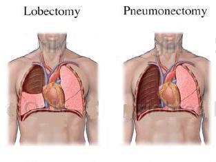
Lobectomy vs Pneumonectomy


The similarities:
1. Both have thoracotomy scars.
2. Both have reduced chest expansion and reduced AE.

The differences:
1. The signs of lobectomy are confined to lobe which is removed. The signs are similar to pleural effusion except it is not stony dull.

2. The signs of pneumonectomy are extensive i.e. involve the whole lung. The side involved would be flatten. It is similar to whole lung collapse.

3. Normally, the tracheal is central in lobectomy except for upper lobe. The tracheal is almost always shifted in pneumonectomy.

Labels:

Friday, March 03, 2006

"Respiratory"

MRCP-PACES 30th November 2005
Station 1:
Respiratory:
A middle-aged lady presented with shortness of breath. Examine her respiratory system. First, I started from the periphery and went up to the head. No abnormality. Then, I went on to feel for the trachea. It was central. As I had discussed with my friend before, I proceeded to examine the back after telling the examiner. There, I noted a needle puncture mark at the left posterior wall. My instinct told me, this should be a case of pleural effusion. It was further confirmed pleural effusion after I elicited stony dullness percussion note with a reduced breath sound and reduce VR. Examiner went on to ask me about how I investigate and manage the patient.

Comment:
This case was relatively straight forward and questions asked was quite direct. I made a terrible mistake here. My friend who sat in UMMC found a centrally placed trachea and he proceeded to examine from the back. After that, he mentioned to the examiner that he would like to complete examination by examining from the front and the examiner said, that was ok and stopped. So, presumed I got the similar scenario, I told the examiner the same thing and he asked me to proceed! I was panicky, there was only three minutes left! Instead, my mind went crazy, I examined the patient's breasts!!! (Thinking there might be a clue there).

Outcome: 4/3 (Thanked God!).

Labels:

Wednesday, March 01, 2006

"Which side first?"

One of the favourite question my friends ask me for respiratory station. It's really depend on what you gather from the peripheral examination namely the tracheal position. If the tracheal is deviated, then I would advise you to concentrate on anterior chest first especially the apical region looking for apical fibrosis or collapse.
If the tracheal is central, then ask the permission to proceed with the back. The differentials would include fibrosing alveolitis, bronchiectasis, pleural effusion or consolidation.

Labels:

Sunday, January 15, 2006

"A scar is a gift"


Sometimes, we panic when we see scars and start thinking of a list of diffenrential diagnoses especially the rare one! But, one of my physican used to calm me down by saying "David, a scar is a gift!"
As in this case with a big thoracotomy scar, one could localize the lesion side by the presence of the scar. Then, you should pay attention to the air entry during auscultation.
a. If air entry is reduced, then think of lobectomy and pneumonectomy.
b. If air entry is equal, then think of decortication and bullectomy.

Labels: